

In addition to preserved specimens, CUP holds over 60,000 photographs as both prints and original negatives. Our images document the last 125 years of plant pathology, mycology, and agricultural practice. Many photographs are tied to specimens in our specimen collection, making them uniquely useful for biological research. Our images include portraits of scientists, agricultural landscapes and apparatus, plant disease symptoms, migrant laborers, pesticide experiments, mushrooms and other fungi, and many other topics. Our earliest photos date back to the 1890s.
Images can be browsed by collection, or searched across all collections through the links below.
We encourage the use of our images in teaching. High resolution files or prints are available for a modest fee. Please preserve our Creative Commons license and provide a link to our site. For other uses, see our terms of use. Lastly, let us know how our images have been useful. Your messages are important in ensuring continued support for this service.
Our photo collections are available to the public via Shared Shelf. You can browse or search them all here:
CUP Photograph Collections (browse and search all images)
You may also navigate our images as individual subcollections — please see below.
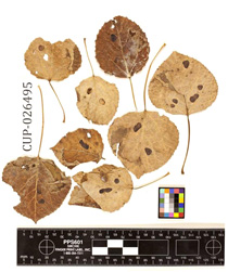
Type specimen of Myrioconium comitatum, CUP 26495.
CUP holds over 8000 types, our most precious specimens. Each is the first of its species to have been described and named, and bears a special nomenclatural significance. So far this collection includes images of over 6000 type specimens at CUP.
Main CUP Photograph Collection
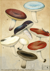
Russula spp. from G.F. Atkinson's Studies of American Fungi (1901). CUP 63522.
Our main CUP collection arises from Cornell's program in Plant Pathology & Plant-Microbe Biology, which was founded in 1907. This is our most diverse image collection, focusing mainly on American agriculture, plant disease, and mycology. It includes portraits of scientists in plant pathology and mycology, and documents the results of scientific experiments conducted by generations of Cornell professors and students.
Main CUP Photograph Collection
G.F. Atkinson Photograph Collection
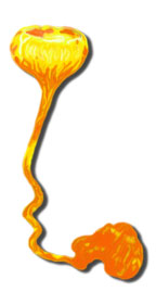
A stinkhorn: Dictyophora duplicata, CUP-A-022858.
The Atkinson Collection includes 8000 images, focusing on mushrooms and other macrofungi that arose from the work of George F. Atkinson, Cornell's professor of mycology between 1890 and 1918. Many of these photographs have associated preserved specimens in our Special Collection CUP-A. You can learn more about Atkinson at our Atkinson website.
Fungi of the Lindsay-Parsons Biodiversity Preserve
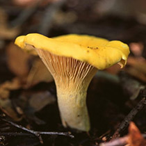
A chanterelle from upstate New York (Cantharellus sp., CUP-LP-026.
The Biodiversity Preserve Collection includes 386 images generated by a bioinventory effort in the late 1990s. It includes images of freshly-collected mushrooms and other macrofungi found at the Lindsay-Parsons Biodiversity Preserve in West Danby, NY. Specimens pictured in these images are vouchered as preserved specimens in our Special Collection CUP-LP.
Robert J. Williams Photograph Collection
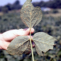
Downy mildew on soybean (1971). RJW-0091.
These 164 color images of plant diseases in tropical agriculture were donated to Cornell by our distinguished alumnus, Dr. Robert J. Williams, who writes:
"The photographs were gathered over many years during the research career of the author, which included more than 13 years as a member of research teams at two of the Institutes of the Consultative Group of International Agricultural Research and 10 years as a research leader with a major multinational agricultural chemical company.
"The collection emphasizes tropical food crops, including cassava, pearl millet, sorghum, rice, maize, and cowpea. It includes: the first ever photographs of cassava bacterial blight and cassava mealy bug in Africa; evidence of the importance of crop-pathogen-environment interactions and the understanding of the biology and epidemiology of diseases; the power of host plant resistance and the key role of effective screening and selection systems in the development of valuable new cultivars; and the effectiveness of simple seed treatments as components of crop disease control systems."
Smokin' Doc Thurston Collection
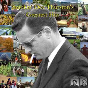
The Smokin' Doc himself: H. David Thurston (1927–2014).
Dr. H. David Thuston contributed almost 2500 photographs from his long career in tropical agriculture. Dr. Thurston was a beloved Cornell professor whose work was in international agriculture, tropical plant pathology, agricultural development in the tropics, sustainable development, and the traditional practices of farmers in developing countries.
The Smokin' Doc's Collection is temporarily offline.
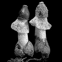
CUP Stereoscopic Photograph Collection
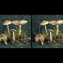
Agrocybe praecox from the Cornell campus, CUP-067587.
Stereoscopic photographs capture some of the three-dimensionality of mushrooms. They can be viewed with a special viewer, but many of us can integrate the two images with our naked eyes on a computer monitor, with a little practice. Our collection includes 53 paired color images of macrofungi.
The Cornell Plant Pathology Herbarium maintains a creative commons license on our images. We encourage the use of CUP images in teaching and not-for-profit education, so long as our identity and license information is prominently included and the Cornell Plant Pathology Herbarium is cited as source. If used on a website, please include a link to our main website.
Arrangement may be made to use our images for other uses (books, greeting cards, websites, scientific publications, and other uses) by consulting the Director.
A word of thanks
Scanning old photographs is not a simple task, and we have many people to thank for their patience, persistence, and participation. Foremost among them is Kent Loeffler, the last in a distinguished line of Plant Pathology Photographers: he trained our interns, combed the collections for interesting images, scanned many, many images, and freely shared his expertise. We also thank photographer Claire E. Smith for all her work, including digital repairs of damaged, century-old glass negatives. Ed Mackillop helped digitize Dr. Thurston's collections, and Prof. Emeritus Wayne Sinclair contributed scanning equipment that facilitated this work. We digitized many of our photos with support from the Anna E. Jenkins fund at Cornell, and also from the National Science Foundation. We also thank the fine folks at Cornell's Olin and Mann libraries for helping us get these images into the public eye.
Places to find our photos
Cornell Mushroom Blog
CUP Photos at Mann Library (an older site presenting a subset of our images)
A collection of preserved plant disease and fungus specimens documenting the world's biodiversity
CUP Pages
CUP Extras
contact:
CUP-herbarium@cornell.edu